Introduction to the Heart
The heart is a vital organ in the circulatory system, responsible for pumping blood throughout the body. It ensures that oxygen and nutrients are delivered to tissues while removing waste products like carbon dioxide. The heart consists of four chambers: two atria (upper chambers) and two ventricles (lower chambers).

Mechanisms of the Heart

To understand how the heart functions, let’s break it down into smaller components:
- Structure of the Heart:
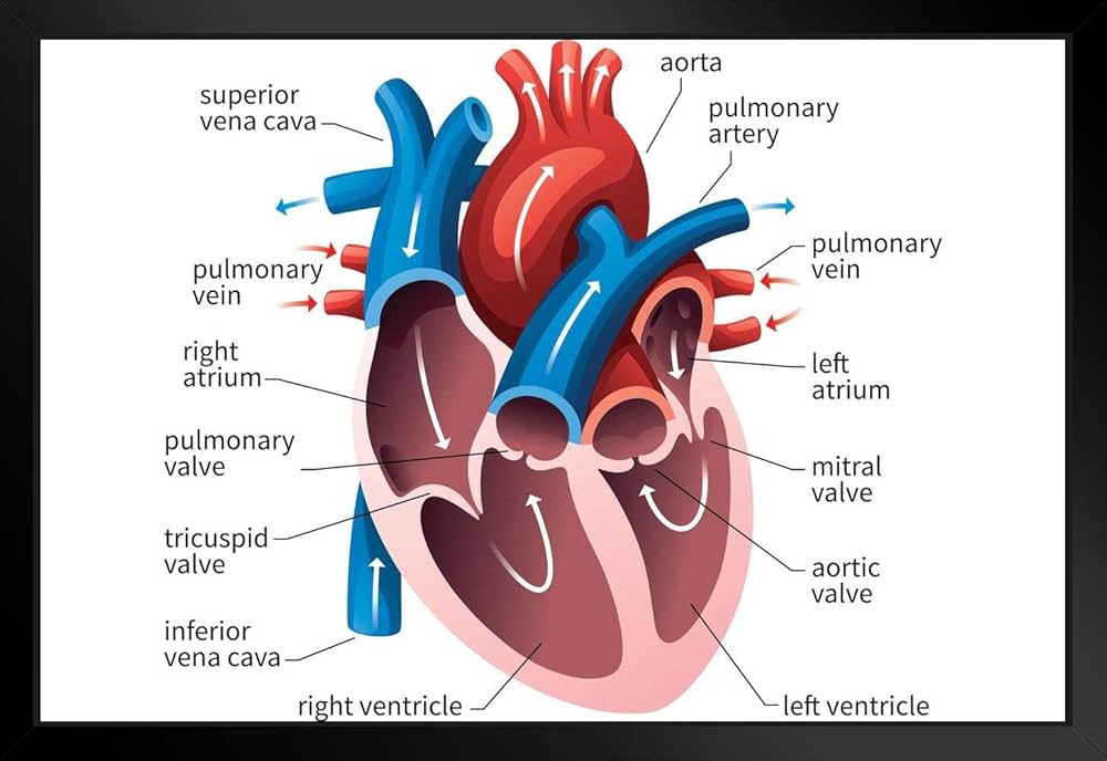
- Chambers: The right atrium receives deoxygenated blood from the body, while the left atrium receives oxygenated blood from the lungs. The right ventricle pumps blood to the lungs for oxygenation, and the left ventricle pumps oxygenated blood to the rest of the body.
- Valves: The heart has four main valves (tricuspid, pulmonary, mitral, and aortic) that ensure one-way blood flow, preventing backflow.
- Blood Flow Pathway:

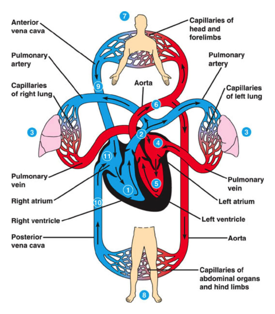
- Blood enters the right atrium from the body via the superior and inferior vena cavae.
- It flows through the tricuspid valve into the right ventricle.
- The right ventricle pumps blood through the pulmonary valve into the pulmonary arteries, leading to the lungs.
- In the lungs, blood gets oxygenated and returns to the left atrium via the pulmonary veins.
- From the left atrium, blood moves through the mitral valve into the left ventricle.
- Finally, the left ventricle pumps blood through the aortic valve into the aorta, distributing oxygenated blood throughout the body.
- Heartbeat Regulation:
- The heart’s rhythm is controlled by electrical signals originating from the sinoatrial (SA) node, known as the heart’s natural pacemaker.
- These signals cause the atria to contract and push blood into the ventricles.
- The signals then travel to the atrioventricular (AV) node, which introduces a slight delay before sending signals to the ventricles, allowing them to fill with blood before they contract.
- Cardiac Cycle:
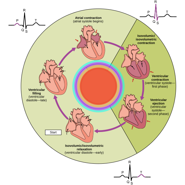
- Phases of the Cardiac Cycle

- Diastole
- Atrial Diastole
- Atria fill with blood from veins.
- AV valves (mitral and tricuspid) are open.
- Ventricular Diastole
- Ventricles fill with blood from atria.
- Semilunar valves (aortic and pulmonary) are closed.
- Atrial Diastole
- Atrial Systole
- Atria contract to push remaining blood into ventricles.
- Ventricles are filled to capacity.
- Ventricular Systole
- Isovolumetric Contraction
- Ventricles contract; pressure rises.
- All valves are closed.
- Ventricular Ejection
- Semilunar valves open.
- Blood is ejected into aorta and pulmonary artery.
- Isovolumetric Contraction
- Return to Diastole
- Ventricles relax; pressure drops.
- Semilunar valves close, preventing backflow.
- Role of the Autonomic Nervous System:
- The autonomic nervous system regulates heart rate. The sympathetic nervous system increases heart rate during stress or physical activity, while the parasympathetic system slows it down during rest.
1. Capillaries
- Structure: Microscopic blood vessels; walls are one cell thick (endothelium).
- Function: Site of exchange between blood and tissues; allow nutrients, gases, and waste products to diffuse.
- Types:
- Continuous capillaries: Found in muscle and brain; have tight junctions.
- Fenestrated capillaries: Found in kidneys and intestines; have pores for increased permeability.
- Sinusoidal capillaries: Found in liver, spleen; larger openings allow larger molecules and cells to pass.
2. Veins
- Structure: Thinner walls than arteries; larger lumen; valves present to prevent backflow.
- Function: Carry deoxygenated blood back to the heart (except pulmonary veins); blood reservoir (can hold up to 70% of total blood volume).
- Types:
- Superficial veins: Located near the surface of the skin.
- Deep veins: Located deeper in the body, often accompanying arteries.
3. Patterns of Circulation
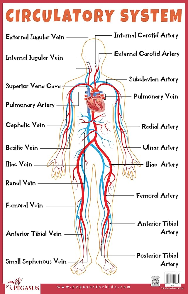
- Coronary Circulation:
- Supplies blood to the heart muscle.
- Coronary arteries branch from the aorta; cardiac veins drain into the coronary sinus.
- Renal Circulation:
- Blood flow to and from the kidneys for filtration and waste removal.
- Heart leaving the blood around 25% to the kidney.
- Renal arteries supply blood; renal veins return filtered blood to the systemic circulation.
- Hepatic Portal Circulation:
- Transports nutrient-rich blood from the gastrointestinal tract and spleen to the liver around 70%.
- Hepatic portal vein carries blood to the liver for processing before returning to the heart via hepatic veins.
4. Blood Pressure
- Definition: The force exerted by circulating blood on the walls of blood vessels.
- Measurement: Typically measured in millimeters of mercury (mmHg) with two readings: systolic (pressure during heartbeats) and diastolic (pressure between beats).
- Normal Range: About 120/80 mmHg is considered normal; variations can indicate health issues.
- Factors Affecting Blood Pressure:
- Cardiac output (volume of blood the heart pumps).
- Blood volume.
- Vascular resistance (narrowing of blood vessels).
- Regulation: Controlled by the autonomic nervous system, hormones (like adrenaline), and kidney function.